12 lead ecg placement
What Is a 12 Lead ECG?
ces 10 physical electrodes to create 12 different views (leads) of the heart’s activity.
This non-invasive test is essential for detecting:
- Heart attacks (myocardial infarction)
- Arrhythmias
- Conduction blocks
- Electrolyte imbalances
- Structural abnormalities
Proper electrode placement is crucial. Even small errors in lead positioning can cause misdiagnosis or false positives on an ECG.
To understand the clinical applications of ECGs, visit the American Heart Association
Why Accurate ECG Lead Placement Matters
Incorrect electrode placement can lead to:
- Misinterpretation of ST-segment elevations or depressions
- Misidentification of axis deviation
- Altered QRS morphology
- Unnecessary treatment or cardiac interventions
For accurate cardiac monitoring, clinicians must know the exact anatomical landmarks for each electrode.
Whether you’re a nurse, EMT, paramedic, or med student—precision matters when it comes to ECG placement.
12 Lead ECG: Basic Components
A standard 12 lead ECG includes:
- 4 Limb Electrodes (RA, LA, RL, LL)
- 6 Chest (Precordial) Electrodes (V1 to V6)
These 10 electrodes create 12 leads:
- 3 Standard limb leads (I, II, III)
- 3 Augmented limb leads (aVR, aVL, aVF)
- 6 Chest leads (V1–V6)
Each lead offers a different electrical view of the heart’s muscle activity.
Limb Electrode Placement (RA, LA, RL, LL)
These electrodes record the heart’s frontal plane activity.
| Electrode | Placement Location |
|---|---|
| RA (Right Arm) | Anywhere between right shoulder and wrist |
| LA (Left Arm) | Anywhere between left shoulder and wrist |
| RL (Right Leg) | Anywhere between right hip and ankle (ground electrode) |
| LL (Left Leg) | Anywhere between left hip and ankle |
Tips:
- Keep limbs still to reduce artifact
- Place electrodes symmetrically for clean tracings
- Avoid bony prominences and use flat surfaces of limbs
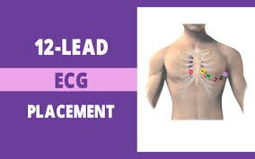
Chest (Precordial) Lead Placement (V1–V6)
These leads record horizontal plane activity of the heart.
🫀 V1
Location: 4th intercostal space, right of the sternum
🫀 V2
Location: 4th intercostal space, left of the sternum
🫀 V3
Location: Midway between V2 and V4
Place after V2 and V4 are in position.
🫀 V4
Location: 5th intercostal space, midclavicular line (left side)
🫀 V5
Location: Same horizontal level as V4, at anterior axillary line
🫀 V6
Location: Same horizontal level as V4, at midaxillary line
Placement Tips:
- Use fingers to find ribs and spaces—do not guess
- Place electrodes under the breast if necessary (especially for female patients)
- Avoid placing over fat, muscle, or bone
To view ECG placement images and learning tools, visit Life in the Fast Lane ECG Library
How to Perform a 12 Lead ECG: Step-by-Step Guide
1. Explain the Procedure
Tell the patient what to expect. Reassure them it’s painless and quick.
2. Position the Patient
- Supine position
- Arms relaxed at sides
- Legs uncrossed
- Ensure comfort to reduce movement artifacts
3. Expose Chest and Limbs
- Provide privacy
- Clean skin (remove oils, lotions)
- Shave if necessary to improve electrode contact
4. Place Limb Electrodes
Apply RA, LA, RL, and LL stickers securely to clean skin.
5. Locate Landmarks for V1–V6
Use anatomical landmarks, not visual estimates.
6. Apply Chest Electrodes
Place V1–V6 in the correct intercostal spaces and lines.
7. Connect the Leads
Attach the ECG machine wires to the correct electrodes.
8. Ask the Patient to Remain Still
Movement or talking can distort readings.
9. Run the ECG
Print or record a 10-second strip once everything is secure.
10. Label and Document
Note date, time, and patient condition during ECG in the record.
Common ECG Placement Errors to Avoid
- Misplacing V1 and V2 too high—often ends up in 3rd intercostal space
- Uneven limb placement—can affect lead I and II
- Loose electrodes—cause artifacts or dropped leads
- Incorrect intercostal counting—always count from the top (start at sternal notch)
Recheck positioning if:
- The ECG shows unusual waveforms
- The axis appears abnormal without clinical cause
- There are missing or inconsistent leads
Special Considerations
🧍♀️ Female Patients
- Always place electrodes under the breast tissue for accurate readings
- Use back of fingers to lift breast if needed
- Maintain professionalism and privacy
🧒 Pediatric Patients
- Use smaller pediatric electrodes
- Adjust placement based on smaller chest size
- Monitor more frequently for motion artifacts
❤️ Dextrocardia (Right-Sided Heart)
- Place precordial leads on the right side of the chest (mirror placement)
🚑 Prehospital ECG (EMS/Paramedics)
- Limb electrodes may be placed on upper arms/thighs if wrists/ankles are inaccessible
- Focus on speed and accuracy, especially in acute chest pain cases
How to Read ECG Lead Views
Each lead gives a unique angle:
| Lead | View |
|---|---|
| I, aVL | Lateral wall |
| II, III, aVF | Inferior wall |
| V1, V2 | Septal wall |
| V3, V4 | Anterior wall |
| V5, V6 | Lateral wall |
Correct placement ensures accurate assessment of each heart region for ischemia, infarction, or conduction abnormalities.
Cleaning and Maintenance
- Use alcohol wipes to clean skin before placement
- Dispose of single-use electrodes properly
- Wipe leads and clips with disinfectant after use
- Inspect cables regularly for damage or corrosion
Final Thoughts: Precision Saves Lives
Proper 12 lead ECG placement is one of the most vital skills in emergency medicine, cardiology, and general healthcare. A well-placed ECG can catch a STEMI, arrhythmia, or electrolyte abnormality before it becomes life-threatening.
Whether in the ER, ambulance, or clinic—accuracy and confidence in lead placement can literally save lives.
Master the landmarks, double-check your work, and never underestimate the power of precision.


